Monday, April 25, 2022

MedTech Review: ELITONE® - Urinary Incontinence Device

PROFESSIONAL EVALUATION OF A URINARY INCONTINENCE DEVICE

By: Dr Roberta Kline (OBGYN)

BACKGROUND
Urinary incontinence is a common problem for women, affecting up to 1 in 3 women over their lifetime. Many treatments have been devised that have varying degrees of invasiveness and effectiveness, with the most utilized approaches including pelvic floor muscle exercises (Kegel’s), surgery and medication. 

Pelvic floor muscle exercises are typically the first-line approach Stress Urinary Incontinence, and while they can often be effective, many women do not know how to do them correctly. This can lead to lack of improvement or potentially create other pelvic floor issues. While pelvic floor muscle exercises can also be helpful for women with Urge Incontinence, they are often are directed toward lifestyle changes and medication. One of the biggest challenges is that many women suffer from both conditions and finding a solution that is effective for both is often elusive. 

But even if a therapy may be effective, there are two large barriers that must be overcome: access to treatment, and then adherence to that treatment. Even if a woman is one of the fewer than 50% that actually seek medical care and get an effective treatment, adherence is often a key reason why it fails to deliver significant or lasting results. Attempts to address these issues are behind the continued innovation in finding effective treatments that are effective, easy to use, and widely accessible.

Electrostimulation of the pelvic floor muscles for treating Urinary Incontinence is not new (Yamanishi et al), but until recently most approaches relied on intravaginal placement of devices. While these have been shown to have good success, utilization rates are typically low due to the invasive nature and inconvenience of such treatments. (Fürst, MCB et al.) For this reason, less invasive ways to deliver electrostimulation technology have been growing in popularity and have been shown to be effective for both types of Urinary Incontinence (Stewart F, Gameiro O et al. and Stewart F, Berghmans B et al.)

Transperineal electrical muscle stimulation (TEMS) is a relatively new and innovative approach utilizing a noninvasive method to treat Urinary Incontinence. With TEMS, electrodes are placed strategically on the perineum so that pelvic floor muscles are stimulated to contract. While more studies need to be done, early clinical research is demonstrating effectiveness (Kim et al,). 

CLINICAL STUDY OF THE ELITONE
In a cohort study published in 2019 (Kolb et al), they found that the women with mild to moderate stress urinary incontinence experienced statistically significant improved symptoms and quality of life scores over only 6 weeks of using the Elitone device. 

I was able to test out and evaluate the Elitone device, which seeks to reduce the disparity in access to effective treatment utilizing this TEMS technology. This is a self-administered device that contains the electrodes in a gel pad that is placed on the perineum, and utilizes electrostimulation to achieve effective contraction of the pelvic floor muscles. The device is easy to use, and the gel pad provides enough adherence without causing discomfort to the skin upon removal. 

As was described in the patient instructions, the TEMS creates a tingling on the skin that can be a little intense at first, but quickly decreases as the nerve fibers in the skin become desensitized. I found the strength of pelvic floor muscle contraction to be quite adequate and not at all uncomfortable, even at high intensity. While the device is designed so that it can be worn during normal activity, I did find that sitting increased the level of contact with the perineum and seemed to increase effectiveness.

As an ObGyn physician, I know that all women with Urinary Incontinence are not alike. They have varying degrees of pelvic muscle floor tone or strength. It’s important to be able to customize the strength of contraction when being stimulated with a TEMS device. Contractions that are too weak for a woman with fairly good pelvic floor muscle tone may be far too strong for one whose pelvic floor muscle tone is much less. This can lead to ineffectiveness on the one end of the spectrum, and pain on the other. Both will lead to lack of the desired outcome of reducing or eliminating the urinary incontinence. 

One of the big benefits is the ability to control the strength of pelvic floor muscle contraction. The device enables easy modulation of the strength of the electrostimulation, thereby enabling a great level of customization for varying degrees of pelvic floor contraction. Over the course of the treatment, each woman can start at the appropriate strength for her, and then gradually increase over time to achieve the level of pelvic floor muscle stimulation needed to maximize strength and thus outcomes. In addition to the impact on pelvic floor muscles, the Elitone device also provides stimulation to the bladder detrusor muscle as a way to address urge urinary incontinence as well – a big plus for the large percentage of women that suffer from both stress and urge symptoms if future studies confirm the effectiveness of this approach.


CONTRIBUTORS

BOBBI KLINE, MD 
(Educational Dir. /Women's Diagnostic Group)
Dr. Kline is a board-certified ObGyn physician, Integrative Personalized Medicine expert, consultant, author, and educator whose mission is to change how we approach health and deliver healthcare. She helped to create the Integrative & Functional Medicine program for a family practice residency, has consulted with Sodexo to implement the first personalized nutrition menu for healthcare facilities, and serves as Education Director for several organizations including the Women’s Diagnostic Health Network, Mommies on a Mission. Learn more at https://bobbiklinemd.com 


References

1) Electrical stimulation with non-implanted devices for stress urinary incontinence in women

2) Fiona Stewart, Bary Berghmans, Kari Bø, Cathryn Ma Glazener Cochrane Database Syst Rev. 2017 Dec 22

3) Stewart F, Gameiro O et al. Electrical stimulation with non‐implanted electrodes for overactive bladder in adults. Cochrane Database of Syst Rev 2021 Dec 9

4) Kim HW et al. Effectiveness of transperineal pelvic floor electrical stimulation system in improving female stress urinary incontinence: a prospective pilot study. Journal of Inclusion Phenomena and Macrocyclic Chemistry volume 101, pages329–336 2021

5) Fürst, MCB et al. Long-term results of a clinical trial comparing isolated vaginal stimulation with combined treatment for women with stress incontinence. Einstein (Sao Paulo). 2014 Apr-Jun; 12(2): 168–174

6) Yamanishi T, Yasuda K. Electrical stimulation for stress incontinence. Int Urogynecol J Pelvic Floor Dysfunct. 1998;9(5):281-90.

7) Kolb G et al. Surface-Applied Electrical Muscle Stimulation for Self-administered Treatment of Female Stress Urinary Incontinence. Journal of Women's Health Physical Therapy: October/December 2019 - Volume 43 - Issue 4

Disclaimer: The information (including, but not limited to text, graphics, images and other material) contained in this article is for informational purposes only. No material on this site is intended to be a substitute for professional medical advice or scientific claims. Furthermore, any/all contributors (both medical and non-medical) featured in this article are presenting only ANECDOTAL findings pertaining to the effects and performance of the products/technologies being reviewed - and are not offering clinical data or medical recommendations in any way. "MEDTECH REVIEWS" is a reporting and publishing project supported by IPHA (Integrated Pain Healers Alliance) and are freely submitted by contributing writers based on their experience(s) of the innovations herein.  These reports are not commercially motivated and are written without any influence by the manufacturer in any way. Always seek the advice of your physician or other qualified health care provider with any questions you may have regarding a medical condition or treatment and before undertaking a new health care regimen, never disregard professional medical advice or delay in seeking it because of something you read on this page, article, blog or website.


Wednesday, April 13, 2022

Smart Sense: Managing Pain Holistically

INTRODUCTION:
Thanks in part to the web-driven information age, today's educated consumer holds the advantage of some of the fastest research technologies and complete global access to the vast majority of health protocols and treatment modalities available.  This forged our personalized culture where optimal info access and the freedom to decide between many diagnostic and therapeutic principles helps us go beyond the TRADITIONAL (or conventional) medicine.  The Integrative Pain Healers Alliance (IPHA) is committed to exploring all areas of health from current experts, researchers and educators.  We start our report with a popular injury called Plantar Fasciitis. We are fortunate to encounter a specialist in HOLISTIC PODIATRY to shed light on a whole-body approach to healing this commonly painful disorder. Dr. Robert Kornfeld offers his successful strategy to fulfilling what many would consider a 'healthier and more intelligent way' to therapy by combining convention with complementary and alternative medicine.


ADDRESSING PLANTAR FASCIITIS HOLISTICALLY

When considering any inflammatory pathology like plantar fasciitis, it is important to not only look at the tissues in pain and make a diagnosis, but it is equally important to understand the mechanisms behind the pathology. In so doing, we need to understand our patient as a unique being and not fall into the trap of treating every patient with plantar fasciitis in the same way. Many patients can develop plantar fasciitis but can have completely different mechanisms that make it difficult for the immune system to heal the injured tissue.

In my world, the world of functional medicine, we need to examine 2 issues before we get into healing the plantar fascia. Number one is we must look at the patient’s BIOMECHANICS, i.e., the structural and functional contributions to the problem. This may be congenital structural issues or problems with the functional aspects of walking and running. It can be poor training, over-training, tight muscles due to lack of stretching, improper footgear for that particular patient’s needs or the necessity to manage these issues with a prescribed, custom molded orthotic device.

Many patients will heal by correcting these biomechanical contributors to the dysfunction which cause the injury to the plantar fascia. But many will not. In those patient’s it become necessary to look at the immune system which is tasked with the job of repairing injured cells. So the second thing we turn our attention to is to discover what burdens there are on the immune system that are making it inefficient and not enabling the healing of the injury. For that, we need to take a patient-specific approach to the epigenetics (diet, lifestyle and environmental issues). The most common things we look for are:

- Poor nutrition/food quality

- Lack of adequate hydration

- Adrenal stress/Neurotransmitter imbalances

- Detoxification disorders

- Hormonal changes

In addition, we must look at what is involved in healing. The plantar fascia, being a connective tissue, receives its blood flow through microscopic blood vessels, not large arteries. In our modern society, we are dealing with many chronic stressors that can lead to adrenal/neurotransmitter imbalance. If there is an excitatory neurotransmitter dominance, we see microscopic blood vessels go into spasm. This is a primitive survival mechanism designed to shunt blood from the extremities and send it to the heart for increased blood flow to the upper arms and legs to either fight the beast that wants to eat us or run away from it. Correcting these chemical imbalances can alter the rate of perfusion into these tiny vessels in order to deliver the necessary substrates of healing.  

HEALING THE PLANTAR FASCIA
Once all of these mechanisms have been diagnosed and addressed (obviously in patients who do not respond to conventional treatments), then we can begin to foster the healing of the tissues. I utilize regenerative medicine which are medicines that stimulate more efficient repair and message the immune system to release protection and open the pathways that can repair the injured cells. This is health promoting instead of using drugs with side effects that interfere with normal physiology.

To that end, I use homeopathic injections, prolotherapy, PRP or amniotic allograft injections. There are different reasons why I choose different approaches but it is all patient-specific. Regardless of the chosen therapy, they are all designed to get the immune activity focused on healing and repair and not on protection. In this way we heal patients AND we leave then healthier than when they first came to see us.


CONTRIBUTOR:
DR. ROBERT KORNFELD has been practicing podiatric medicine and surgery for over 40 years. After training in traditional medicine and surgery, Dr. Kornfeld sought to answer a very important question - why does this specific patient have this diagnosis? He knew that finding the underlying mechanism to the presenting pathology was the critical issue that traditional medicine was not addressing. This naturally brought him into the field of holistic medicine. His dedication to treating chronic foot and ankle pain at its source has given him well-researched insight into how the human body manages cellular injury that it is unable to repair so he can alleviate chronic pain syndromes that have resisted traditional medical approaches. Dr. Kornfeld has offices in New York City and Port Washington, Long Island. (https://www.drrobertkornfeld.com/)


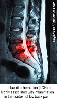
The research benefits of using ultrasound scanning in therapeutic monitoring allows for tremendous flexibility in exploratory detection. Once we have established the initial Base Line of the ‘before and after’ scan, having probe in hand empowers the diagnostician to go deeper and wider- allowing for more answers and possibly finding other pathologies.   After we looked at the obvious muscle abnormality, the question is to go back and see what's causing the problem. Upon observing what’s under the white skin area, there appears a very thick white band (FASCIA covering the muscle) between the two dark layers of the subcutaneous fat and the muscle.  We notice that the fascia splits into two levels with the upper arrows, denoting the top part of the split and the lower arrows showing the bottom part of the split. The black area in the split is filled with fluid indicating the fascia lining is abnormal and this is a way that we can follow the cause for the muscle irritation in future tracking.  (see complete article & video)


PAIN 101: PLANTAR FASCIITIS BREAKDOWN 
by: Josh Tiberius Schueller, PT

Plantar Fasciitis is a challenging, difficult and often time frustrating injury that effects millions of people per year. PF is not considered a serious injury on the spectrum of musculoskeletal injuries so there have been a lack of attention to effective treatments. Many suffers are active competitive people who struggle with the lack of effective treatment. These people have to take weeks into months off from training because of this injury. Traditional treatments have been lackluster in results which has spawned “gimmicky” devices that have flooded the market. The need for a safe, effective treatment for PF is apparent. These devices need to be validated to decrease consumer fraud. The health care professionals need to realize that traditional treatments of ice, rest, and immobilizing are not effective for active people. (see full article)


Wednesday, March 30, 2022

DOG INTERACTIVITY: A HORMONE STIMULANT FOR OUR MENTAL HEALTH

INTRODUCTION
The IPHA Health News is proud to launch our spring edition with this scientific review on how expressing love for 'man's best friend' actually works as a measurable therapeutic activity.  Brought to you by our clinical and mental health researcher, Dr. Bobbi Kline, this unique feature explains the physiological response between our touching or connecting with a dog and a biometric reaction to our endocrine system.


THE HEALING POWER OF PAW-SITIVITY 
Written by: Dr. Bobbi Kline

The relationship between dogs and humans began since the beginning of time when dogs looked to humans as a means of survival. Numerous studies have reported the significant benefits of pet ownership and interactive coexistence -  from stress reduction to social fulfilment to a healthier lifestyle.  Science is now providing insights into how this works, and the two hormones that are responsible: OXYTOCIN and CORTISOL.  A landmark study in 2015 revealed how dogs and their owners emotionally bond, and it’s related to the production of oxytocin – also known as the bonding hormone. When released, oxytocin creates a powerful social attachment between one person and another and creates that “feel good” sense of nurturing and closeness. [1]


THE SCIENCE OF THE MAGNETIC GAZE
It is known that a lengthy eye contact (or mutual gaze) forges a unqiue engagement between two individuals that somehow activates the release of our oxytocin, which also mediates a powerful emotional bond. One of the best-known examples of this is in the bond created between mother and infant. The oxytocin response is a feedback loop- a nurturing gaze from mom releases oxytocin in the infant, which stimulates production of oxytocin in the mother, then further stimulating nurturing behavior into a self-sustaining positive feedback loop. 

This study demonstrated that the same process happens when a dog and owner engage in the same behavior. For the first time, this oxytocin-mediated bonding has been shown to occur outside of intraspecies relationships.  A prolonged eye contact between dog-to-owner has shown to produce an oxytocin response in both the dog and the human owner. Unlike wolves, or even primates who are genetically closer to humans, dogs alone are able to elicit this oxytocin response loop. This may explain the extraordinary bonds formed between dogs and our human owners, and why puppies often exhibit similar behavior patterns as young children in response to visual cues. It may also explain why many dogs grieve when separated from their owners.

Oxytocin also has been shown to reduce stress and increase social reward behavior. But when people have SNPs or small errors in their genes involved in this pathway – including the receptor for the hormone oxytocin – they don’t always get the same level of benefit. [insert link to genetics vs genomics article]  Often they need more oxytocin than they get from a short interaction – something that can be challenging in today’s busy world. Perhaps this is one of the mechanisms behind the success of service dogs for people with disorders involving impaired social pathways, such as autism, PTSD, and other neurodevelopmental conditions where oxytocin is being used as an experimental treatment.

PHYSICAL TOUCH
But it may not only be about oxytocin. More recent studies have added a second hormone into the equation: CORTISOL. [2] Known as the stress hormone, cortisol is released as part of the normal stress response. It is part of the “fight or flight” reaction that helps us flee from danger or hold our ground to confront it.  The challenge is that so often in our modern world, that stress response doesn’t go away when the threat does. Our brains and nervous system stay on high alert far longer than needed, resulting in chronic stress which wreaks havoc with our biology. 

While oxytocin is produced through the gaze, physical touch is the secret behind the stress-reducing power of dogs. When we hold or pet a dog, especially our own but this is not as critical an element as with the oxytocin, science has shown that cortisol levels go down. This, in turn, reduces the “fight or flight” stress response and gives us a chance to reset those alarm bells – and our biologic response. 

As with oxytocin, there are genes involved in the cortisol response that when they have these small errors, or SNPs,  can predispose a person to a heightened or prolonged stress response. This can be a significant reason to why the same experience that one person brushes off can lead to serious consequences in another – including anxiety, depression, PTSD, and even many chronic health issues thought to be purely physical – heart disease, obesity, autoimmune disease, and cancer just to name a few.

So when dog owner says they feel better around their dog, we now know why.  One can venture that a key reason behind the healing power of dogs is the combination of increasing the oxytocin response and reducing the cortisol-driven stress response. It’s science, it’s biology, and it’s their unconditional love that makes this partnership between dogs and humans so enduring.


REFERENCES

(1) Nagasawa, M et al Oxytocin-gaze positive loop and the coevolution of human-dog bonds. Science 17 April 2015: Vol. 348 no. 6232

(2) Petersson M et al.  Oxytocin and Cortisol Levels in Dog Owners and Their Dogs Are Associated with Behavioral Patterns: An Exploratory Study


Monday, February 14, 2022

MITOCHONDRIA EXPLAINED VIA CHRONIC FATIGUE

MedScienceNews
Review on CFS/ME (Chronic Fatigue Syndrome and Myalgic Encephalomyelitis

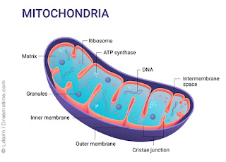
One of the key underlying findings in conditions causing chronic mental or physical fatigue is dysfunction of the mitochondria. Mitochondria are small structures located in the nucleus of every cell, and every cell contains thousands of them. These powerhouses produce the energy our bodies need to carry out every function in the form of ATP (adenosine triphosphate). When mitochondria don’t function at their best, or too many of them are destroyed, our energy levels suffer. While this can be due to inherited genetic disorders, most often it is seen as the result of chronic damage over time.

Many of the biochemical reactions in our bodies produce toxic versions of oxygen, hydrogen and nitrogen, including how we make ATP. These toxic molecules, which we call free radicals, have to be neutralized so they don’t damage the mitochondria. Our bodies have powerful antioxidant defenses to keep these in check. But when these protective systems become overwhelmed by too many free radicals, oxidative stress results and mitochondria are damaged.

Each person’s ability to handle these toxins and oxidative stress is different and depends on the interaction between their genes and their environment. Our genes commonly have small errors, called SNPs (single nucleotide polymorphisms) that affect how each protein in our body works. [see: genetics vs genomics article] Since proteins run all of our biochemistry, these small DNA changes can affect how we deal with emotional stress, energetic stress, and physical stress; how we process the toxins; what we need nutritionally. When we get a mismatch of our genes and our diet and environment, that can set us on a collision course for disease if it persists over time.

Some of the most common causes of oxidative include deficiencies of key nutrients and antioxidants (such as CoQ10, Alpha-lipoic acid, B vitamins, and Vitamin C), and overload of toxic substances in our environment (including air pollution, heavy metals like mercury and arsenic, herbicides and pesticides). Emotional stress is also often a key factor. Viral and other infections that trigger an immune response that then gets out of hand is also a common contributor. Chronic inflammation goes hand -in-hand with the oxidative stress, and this is why so many chronic diseases related to inflammation are also linked to mitochondrial dysfunction – either as a cause or as a result. Once this cascade of oxidative stress and inflammation gets going, it feeds itself and can be hard to reverse unless all contributing factors are addressed.

Because mitochondria are found in every cell of the body, often multiple systems are affected. This can result in a wide range of symptoms that are often hard to diagnose, especially in conventional medicine. Fortunately, we have ways to understand what each person needs through personalized approaches. By integrating functional medicine, genomics, and a wide range of healing modalities, we have the knowledge and the tools to unlock what each person needs to restore health – and then keep them healthy. 


Dr. Bobbi Kline combines her decades of experience as an ObGyn physician, energetic healer, functional and genomic medicine practitioner, along with Human Design and Astrology to help women get to the root of the health of mind, body, and spirit. She is dedicated to serving her fellow clinicians in addition to her patients, she has created and taught courses in functional, genomic, energetic medicine within a holistic model through faculty positions with the University of New England College of Osteopathic Medicine and The Graduate Institute. Currently, as founder of bobbiklinemd.com, she shares the wisdom gained from her own health journey to empower women in successfully navigating life and health challenges for themselves and their children.


SPOTLIGHT SERIES:

Climbing Out of CFS with Alternative Therapy


SHOT DOWN BY CFS
In 2017, Suzanne Wheeler was diagnosed with Myalgic Encephalitis or Chronic Fatigue Syndrome. She was home bound for about two years and bed bound about 50% of the time. Suffering from significant  chronic pain throughout her body with a health report reading equivalent to that of about a 95 year old, Suzanne spent the better part of 15 years being mis-prescribed therapeutic drugs and dangerous opioids that gave little or no positive results. Read Suzanne's exploratory journey to wellness, her extensive research and her ultimate solution to getting back on her feet again with PEMF. 


Our Friends and Supporters:


Disclaimer: The information (including, but not limited to text, graphics, images and other material) contained in this article is for informational purposes only. No material on this site is intended to be a substitute for professional medical advice or scientific claims. Furthermore, any/all contributors (both medical and non-medical) featured in this article are presenting only ANECDOTAL findings pertaining to the effects and performance of the products/technologies being reviewed - and are not offering clinical data or medical recommendations in any way. Always seek the advice of your physician or other qualified health care provider with any questions you may have regarding a medical condition or treatment and before undertaking a new health care regimen, never disregard professional medical advice or delay in seeking it because of something you read on this page, article, blog or website.


Thursday, February 10, 2022

BREAKING THE CHAINS OF A SITTING LIFESTYLE

Written by: Josh Schueller, PT

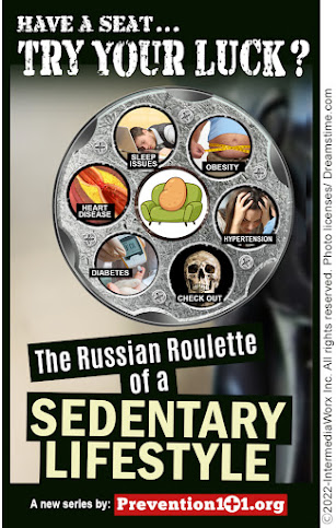
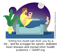
In the health care universe, clinicians face many challenges in the pursuit of improving health and wellness for their patients. Many variables play a role in a patient’s recovery from illness or injury. Some of these variables are controllable some are uncontrollable.  In my decades or treating illness and injury, the #1 controllable variable is having a SEDENTARY LIFESTYLE.   This lifestyle is characterized by a high rate of inactivity (such as too much sitting) and little physical activity throughout each day-- a major precursor to many serious health issues.  

CONVENIENCE AND COMFORT: KILLER HABITS FROM TECHNOLOGY
The past 20 years 'blessed' humanity with an exodus of screen technologies like phones, computers, and streaming videos on TV- increases the prevalence of sitting stationery.  The average American spends over 7 hours a day looking at a screen of some kind. Combining this with 7-9 hours of sleeping per day, two-thirds of our day is automatically spent sedentary for the average American.  This doesn’t account for time spent driving, eating, or talking with someone which usually occurs in a sitting position.  Many have compared sitting as the new smoking regarding health concerns. Nearly a half of all Americans are obese.  Sedentary lifestyles are directly correlated with obesity.  Hypertension, heart disease, Type 2 diabetes, colon cancer, and sleep apnea are all significantly increased because of a sedentary lifestyle.  Musculoskeletal issues such as osteoporosis, joint pain, chronic inflammation and stiff/weak muscles are all associated with sedentary lifestyle. Anxiety and depression are increased with sedentary lifestyle.  


Over the last two years COVID-19 has also played a significant role in increasing sedentary lifestyle.  Social isolation, social distancing, working from home, fitness centers closing, quarantine have all led to an increased acceptance to a sedentary lifestyle.  Over the last two many of our houses have a worn carpet trail from the bedroom to home office to kitchen to bathroom every single day.  Rinse and repeat every day.  Blaming COVID is a popular rationale. A recent study from the British Journal of Sports Medicine, demonstrated that sedentary people had a greater risk of hospitalization, admission to ICU, and death from COVID-19 compared to those who meet the physical activity guild lines. 


THE WRONG WAY TO TRY
Looking at our current existence (job types, transportation modes, technologies), modern society has enabled us to be inactive.  In fact, it encourages it.  Our very economic trends like online ordering lends itself to us all reaching for the quicker/cheaper option instead of exerting any calories to achieve anything. 

Exercise is the new swear word in today’s world.  Many people just hearing the word exercise break out in hives or have a breakdown.  The excuses not to exercise are endless.  “I am too busy”, “I am too tired”, “it hurts to exercise”, “exercise is boring” seem to be the most common.  We need to stop using the words exercise and diet and instead replace with movement and wellness.  So many people take a failed approach to movement and wellness.  This “all or nothing” mind set has to stop and it has to stop NOW!!!!.  If you have been sedentary for an extended period, DO NOT try to change a hundred bad habits.  That is unfair and it leads to failure.  Set yourself up for success not failure.  Success breeds success.  If you think you are going to change all your bad habits like physical activity, diet, sleep, and medications all in one day or week you find yourself back on the couch watching Real Housewives of Whatever County.

According to just about every cardiologist and Fitbit(tm), movement is recommended 150 minutes a week. That might seem like a lot but that is only 30 minutes a day 5 days a week.  That doesn’t mean a high intensity Navy Seals style workout session.  It is easy to fall out of shape, don’t set too many obstacles in your way. 


UPCOMING FEATURES


REFERENCES:
1) Image insert: "Sitting too much can AGE you by a lot..." - AARP: https://www.aarp.org/health/healthy-living/info-2017/sitting-too-much-can-age-you-a-lot-se.html

Wednesday, February 9, 2022

TROUBLESHOOTING MYOCARDITIS


 By Bobbi Kline, MD (Integrative Physician / Genomic Research Specialist)

As a mom, my heart grieves for parents, including Elysa’s, who suffer such devastating tragedy. It’s the worst thing you hope never happens to your child, and I truly admire parents who turn a tragedy into something positive. It requires such amazing strength, courage and grace. As a physician, I find my self immediately asking "Why do these things happen? How can we predict or prevent them?" As clinicians, we look for patterns to help guide diagnosis and treatment. We know what to expect, but sometimes they can lull us into a false sense of security. Childhood viruses, as any parent knows, are an expected part of those early years. 

But what happens when they turn out to be something more? That’s where pattern recognition is crucial. When something obviously falls outside those patterns, it’s a signal to question and go deeper. But what happens when you don’t even recognize that deviation? What if something is so uncommon or so subtle that it’s hard to detect among all the noise? Post-viral myocarditis is one of those conditions, and I’m glad to see it now in the spotlight. Raising awareness is a key first step. While COVID-19 has certainly helped to highlight this condition, it goes further than COVID. Many common childhood viruses have been implicated in causing myocarditis, but most people are completely unaware. I admit that it was not something I ever really thought about as my kids were growing up. And I am not alone. Educating clinicians as well as parents on what to look for, when to be alarmed, when to go deeper is crucial. This alone will save lives. 

But it’s only the first step.
We also need better tests and tools to quickly and easily identify who is at risk, and better treatments for helping these children. This requires a multidisciplinary approach that includes better diagnostics including noninvasive technology, along with effective medications and other treatments. It also includes the burgeoning field of genomics and personalized medicine, both to provide a better understanding of the why, as well as a powerful tool to predict and prevent. For, at the heart of this, is understanding each child’s uniqueness in a way that empowers.  Two studies have been published this year that have the potential to leverage the power of DNA to identify who is at risk for developing myocarditis after a viral infection. Not only that, but also which of those children are most likely to recover, and therefore need fewer interventions, and which of those children are most at risk for sudden death and require much more intensive treatment and support. And, in today’s world, we also need the power of legislation to make sure everyone has access to this higher level of care. There is much promise to change the trajectory of this devastating illness, and it is only through advocacy such as this that it will happen. 


PEDIATRIC MYOCARDITIS & NATIONAL DETECTION MOVEMENT (Part 1)

Written by: Dr. Robert L. Bard

Since the advent of Covid-19 Long Haul studies in 2021, the medical diagnostic community shifted into overdrive- seeking out all available screening and examination protocols to assess health problems called POST-ACUTE SEQUELAE (PASC). One of the recent Covid-19 related headliners is the rise in cases of MYOCARDITIS in children 16 years and under. CDC Reports link the pathological impact with covid infection since it is proven that Viral infections are a common cause of myocarditis. 

Between early 2020–2021, patients with Covid-19 had nearly 16 times the risk for myocarditis[1]. According to the CDC, in a study of myocarditis cases, 2,116 (41.7%) had a history of Covid-19.  In addition, cases of myocarditis reported to the Vaccine Adverse Event Reporting System (VAERS) indicated links between Myocarditis and Pericarditis to come from the mRNA Covid-19 vaccination (especially in male adolescents and young adults) more often after the second dose.[2].


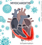
Myocarditis is defined as an inflammatory disorder of the heart muscle (myocardium) leading to cardiac dysfunction. It is also recognized as myocardial cell death 
[3]. Checkups for this also reviews for PERICARDITIS (the inflammation of the outer lining of the heart). Various causes of myocarditis includes: Viral Infectious including adenoviruses, echoviruses, enteroviruses like the coxsackie viruses. In addition, predisposition can occur from those with Autoimmune diseases such as Celiac disease, Churg-Strauss syndrome, Crohn disease, Kawasaki disease, lupus, rheumatoid arthritis, sarcoidosis etc. (See NIH chart for full list of causes- [4])

STANDARDIZED DIAGNOSTICS
As with any critical disorder, detecting early stages of myocarditis allows for a higher opportunity to treat and even eliminate the health risk.  In children, symptoms include: Fever, Fainting, Breathing difficulties, Rapid breathing, Chest pain and Rapid or irregular heart rhythms [5]. In adults, symptoms range from chest pain, shortness of breath, at rest or during activity and fluid buildup with swelling of the legs, ankles and feet. To prevent possible heart damage, a cardiologist may order one of a number of imaging options:

▪ Electrocardiogram (ECG or EKG)
▪ Chest X-Ray
▪ Heart MRI
▪ Blood Tests
▪ Doppler Ultrasound for Acute Myocarditis
▪ Cardiovascular MR Elastography (MRE)
▪ Ultrasound Elastography

See expanded details on diagnostic protocols, visit:  http://pediatricscan.com/myocarditis.html


NEW GROUP: MEET MOMMIES ON A MISSION! 
THE 2022 NATIONAL "ONE VOICE" FOR CHILDREN COLLECTIVE
In Feb. 2022, lead directors of The AngioFoundation, The NY Cancer Resource Alliance and publishers of PREVENTION101.org forged a collaborative outreach community project with the For Elysa Foundation (myocarditis awareness org) to form a national coalition of clinical support associations, research foundations, researchers and parent groups dedicated to the awareness of rare pediatric diseases and critical disorders. This collective concept called "MOMMIES ON A MISSION" is a unique and remarkable platform that aspires to support the many pediatric disorders that plague today's children.

Known as 'Project One", Mommies on a Mission will launch the ONE VOICE crusade to leverage new awareness response for the deadly health threat of Myocarditis in America's children. "MOMMIES ON A MISSION" is a creative title Mrs. Jana Rojas (director of For Elysa Foundation) used to describe her fellow advocates who, like her, established a dedicated benevolent program to battle pediatric myocarditis through education, awareness and to support clinical research. Even more are driven to forge standardized screening of myocarditis as part of a national prevention and early detection initiative. "Losing another child to this terrible and insidious disease is one child too many! It's time we joined hands with all groups, legislators, medical societies and anyone who can make a difference. We hope to deploy a multi-stage battle plan to address this disease and enact new solutions in every pediatrician's office!", states Dr. Noelle Cutter (Molloy College, NY Cancer Resource Alliance).


MedNews Extra
Saving Lives Through Advocacy & Research:
FOR ELYSA FOUNDATION

Around the first week of Feb, 2022, our clinical diagnostic researcher, Dr. Robert Bard launched his PediatricScan.com 2.0 in NYC‐ which included a Pilot program for Myocarditis Screening through the use of advanced Doppler Ultrasound Imaging.  To establish the clinical network for this program is to connect with ICU specialists & Cardiologists as well as all associations supporting Myocarditis research.

We met the directors of a remarkable national advocacy foundation called FOR ELYSA FOUNDATION‐ a non‐profit organization dedicated to promoting Education, providing Light, and supporting Research in the areas of viral myocarditis and pediatric sudden cardiac arrest. (www.ForElysa.org). Mrs. Jana Rojas and husband Jaime Rojas from Kansas City developed this organization inspired by the loss of her vibrant little girl,  Elysa Louise Rojas who passed away at the tender age of two years old. " In Elysa’s case, a common childhood virus was responsible for her myocarditis. The virus either attacked Elysa’s heart directly or caused her immune system to attack her heart muscle in a “friendly fire” fashion while trying to fight the virus. The inflammation in her heart increased drastically and very quickly to the point of sudden cardiac arrest. Doctors and scientists do not fully understand the mechanisms within the body that cause a virus to “go haywire” in the immune systems of individuals with myocarditis. There is currently no way to predict when/if this will occur."


The FOR ELYSA FOUNDATION is one of our first advocacy friends in pursuit of bringing national awareness and supportive clinical research for myocarditis diagnostics and prevention. According to the ForeElysa.org website, Myocarditis is a disease marked by inflammation and damage of the heart muscle. There are many causes of myocarditis, including viral infections, autoimmune diseases, environmental toxins, and adverse reactions to medications. The most common cause of myocarditis in North America is viral infections. Myocarditis usually attacks otherwise healthy people. It is believed that 5 to 20% of all cases of sudden death in young adults are due to myocarditis. Although the exact incidence of myocarditis is not known, it is estimated that approximately 343,000 people die of myocarditis and its major complication, cardiomyopathy, each year. The prognosis is variable but chronic heart failure is the major long term complication. Myocarditis and the associated disorder of idiopathic dilated cardiomyopathy are the cause of approximately 45% of heart transplants in the United States.  

Materials in this excerpt are published with express consent from The For Elysa Foundation.  For complete Information, visit www.FORELYSA.org


HOW & WHEN TO CHECK FOR PEDIATRIC MYOCARDITIS
By: Jana Rojas
The tricky thing with myocarditis being virally mediated is that Elysa could have had a heart scan a week before she died (the day before she contracted the virus that wrecked her heart), and it would have been normal. I am hesitant to insinuate that imaging could "clear" a patient and provide a clean bill of health without noting that this can and does occur spontaneously after viral infections, and so testing while ill or post-virally is actually the key message and window of opportunity for myocarditis detection.
In my mind, the primary role for cardiac diagnostic imaging as it related to myocarditis specifically would be for: 
1) acutely ill children in ED/urgent care/hospital inpatient settings
2) children exhibiting the signs and symptoms you have outlined (fainting, sudden fatigue, shortness of breath, chest pain, palpitations), 
3) after known Covid or other viral infection with prolonged or delayed healing (ie ongoing fatigue, shortness of breath, etc)  
4) and possibly PRE-PARTICIPATION SPORTS PHYSICALS. The pre participation screenings would be enhanced cardiac screenings in general to ideally pick up congenital heart defects and other concerns as well as myocarditis. 


1) Morbidity and Mortality Weekly Report (MMWR): Association Between Covid-19 and Myocarditis Using Hospital-based Admin Data 3/2020-1/2021) https://www.cdc.gov/mmwr/volumes/70/wr/mm7035e5.htm
3) MR Imaging of Myocardial Infarction | RSNA-Radiological Society of North America  / https://pubs.rsna.org/doi/10.1148/rg.335125722
4) The Diagnostic and Clinical Approach to Pediatric Myocarditis: A Review of the Current Literature (NCBI/NIH)  https://www.ncbi.nlm.nih.gov/pmc/articles/PMC6352488/
5) Diagnosis and Management of Myocarditis in Children (American Coll. of Cardiology) https://www.acc.org/latest-in-cardiology/ten-points-to-remember/2021/07/09/17/31/diagnosis-and-management-of-myocarditis

Disclaimer: The information (including, but not limited to text, graphics, images and other material) contained in this article is for informational purposes only. No material on this site is intended to be a substitute for professional medical advice or scientific claims. Furthermore, any/all contributors (both medical and non-medical) featured in this article are presenting only ANECDOTAL findings pertaining to the effects and performance of the products/technologies being reviewed - and are not offering clinical data or medical recommendations in any way. Always seek the advice of your physician or other qualified health care provider with any questions you may have regarding a medical condition or treatment and before undertaking a new health care regimen, never disregard professional medical advice or delay in seeking it because of something you read on this page, article, blog or website. None of the information provided should be interpreted to be or is meant to be medical advice, suggestions, or counseling.

FIGHTING RECURRENCE WITH POSTOP SCANNING

FOR IMMEDIATE RELEASE: 8/6/2024- Dr. Robert Bard (Bard Diagnostic Imaging) and Cheri Ambrose (President of the MBCGA / Male Breast Cancer G...