
One of the most exciting opportunities in neuroscience research today is the use of strategies that protect the brain which may potentially prevent, delay or inhibit the progression of neurodegenerative diseases such as Alzheimer’s, Parkinson’s and ALS. This opportunity rests on our ability for early diagnosis. Research has shown that the likelihood of success for a given treatment-whether lifestyle changes or pharmacological approaches- is highly dependent upon early intervention, before the disease process has become too severe and potentially irreversible. Therefore, it is critical to understand the underlying biological changes that occur in the early stages of these diseases, along with the technology to detect them. *Visit this complete article @ HEALTH SOURCE DIGEST
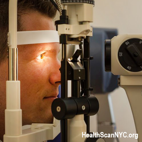
 While ultrasound of the eye has been around for half a century, one of the quick things we can do is find out eye cancer, specifically melanoma in the back of the eye without dilating the eye. Instead of a three hour examination with drops that blur your vision, use of ultrasound scanning allows us to detect an eye cancer in minutes. In a recent report by the American Academy of Ophthalmology, eye exams are recognized to find links to a growing array of diseases. A surprising list of conditions include:
 | • Aneurysm | • Brain tumor | • Hypertension | • Ischemic Stroke |
| • Heart disease | • Lyme disease | • Rheumatoid Arthritis | • Cancers of blood, tissue or skin |
| • Diabetes | • STD's such as Syphilis, herpes, chlamydia, HIV, gonorrhea, genital warts and pubic lice | ||
Though further testing is required once these symptoms may arise during an exam, current diagnostic imaging scans reflect advancements in analysis of the eye in relation to the patient's physiology. [1]
TRENDING: PATHOLOGY LINK OF CHRONIC DISORDERS THROUGH THE EYE
By: Dr. Robert L. Bard

In 2009, the radiological community started investigating the systemic disease of inflammatory arthritis as it manifested in the eye and in the joints. Specifically, people with back pain were getting x-rays, which showed nothing. Based on the poor response of an x-ray and back pain correlation, we started looking at ultrasound and MRI and we found two things; the MRI was much more sensitive than standard back x-rays and the blood flow technology of the Doppler was even more sensitive in showing inflammatory changes in the joint. This is important because (est 2010) a prominent Rheumatology publication discussed inflammation of the Iris (called IRITIS) and this is where the manifestations of Sacroiliitis or ankylosing spondylitis are found. These are signs of inflammatory disease, which affects not only the bones and the joints, but they also affect the eye.
 |
DETECTION OF SYSTEMIC DISEASE
We find the eye to be a pathological mirror into many conditions in the body, such as metastatic disease going to the brain and causing swelling of the optic nerve. More interesting is the correlation between inflammatory disease and eye symptoms. Rarely if ever does a knee surgeon ask about a patient’s eye pain or an orthopedic back surgeon ask about changes in the eye. But significant evidence targets the eye as a source of guidance to where else the disease might be occurring and what the disease actually is.
Specifically in inflammatory back pain or inflammatory arthritis of the joints, we see inflammatory changes in the front of the eye. The same pathologic effect happens with other common dermal inflammatory diseases. Specifically, 6% of the population worldwide has psoriatic arthritis, which also shows up in the eye. 3% of the population has rosacea, which gives you redness in the face and redness in the eye and itching in the eye, but also is a cause of arthritis.
Other areas that we can look at are GLAUCOMA. Through imaging technologies, we see increased pressure in the eye as decreased blood flow. Similarly, increased brain swelling is reflected by a decrease in blood flow to the eye artery and increased pressure inside the eyeball.

In DIABETIC RETINOPATHY, because the inflammatory changes in the vessels have altered the normal velocity of the blood, we are able to use the blood flow feature of the Doppler ultrasound to assess the vessels going to the back of the eye, including the almost microscopic vessels in the retina. This is important because changes from vascular diseases such as eye disease, heart disease, high blood pressure and high cholesterol are all demonstrated in the blood flow to the back of the retinal area. This technology is also widely performed with other optical technologies such as optical coherence tomography, commonly used by ophthalmologists and dermatologists. However, the Doppler blood flow was not recognized by the American ophthalmologic community as a safe tool until recently.
Another cause of eye disease and migraine is called GIANT CELL ARTHERITIS- an inflammatory disease, which affects the blood vessels going to the area of the eye. The standard diagnostic treatment is to cut out the painful artery (biopsy it) and see if it's diseased. However, today we put the ultrasound probe on the temporal artery, which goes to the outside of the eye. Oftentimes the patient will take the probe and put it on the painful side, representing the greatest area of inflammation and will hold it for us as we turn on the blood flow technology. This shows increased flow in the area that's narrowed and soft tissue swelling of the outer vessel wall, demonstrable on ultrasound in seconds.
Because ultrasound is a real time tool that shows blood flow as a treatment is being done, (such as with electromagnetic stimulation) we are able to visually confirm noninvasively the impact of treatments on the arteries that go to the retina and choroid and notice changes such as dilation with some of the new treatments that are available. Hopefully this will bring relief to sufferers of diabetes, glaucoma, and Alzheimer's disease. Since this noninvasive diagnostic tool is safe, repeatable and quantifiable.
PART 2: SCANNING COGNITIVE DECLINE

This case subject with a presumptive degenerative neuromuscular disease or amyotrophic lateral sclerosis. In a non-invasive investigation, we look at the eye for increased intracranial pressure, which may reflect in changes in the optic nerve diameter. [Fig. 1] The right eye, which is the left slide, shows the optic nerve diameter of five millimeters. And the right is almost eight millimeters. So we have a guide as to where the brain disease is more severe. At this point, we activate the blood flow function and look at the right eye and see that the blood flow from the anterior cerebral artery circulation that supplies the back of the eye is approximately 60 per second in the blood flow which corresponds to the graph on the bottom. [Fig. 2] On the left eye. The same blood vessel shows a decreased pressure of approximately 45 as shown by the decreased height of the blood flow graph at the bottom. Once again, we have measurable diagnostic technologies that are noninvasive, so we can follow treatment in diseases that are affecting the eye and as it relates to brain and degenerative neuromuscular disorders. (See complete feature on Neurological Studies)


Disclaimer: The information (including, but not limited to text, graphics, images and other material) contained in this article is for informational purposes only. No material on this site is intended to be a substitute for professional medical advice or scientific claims. Furthermore, any/all contributors (both medical and non-medical) featured in this article are presenting only ANECDOTAL findings pertaining to the effects and performance of the products/technologies being reviewed - and are not offering clinical data or medical recommendations in any way. Always seek the advice of your physician or other qualified health care provider with any questions you may have regarding a medical condition or treatment and before undertaking a new health care regimen, never disregard professional medical advice or delay in seeking it because of something you read on this page, article, blog or website.